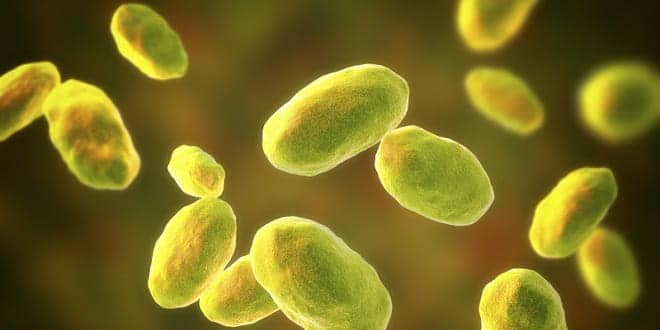
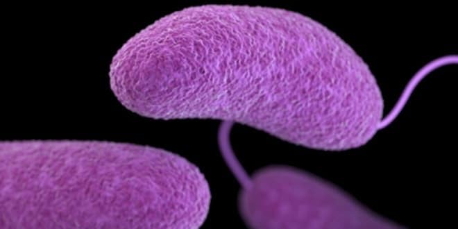
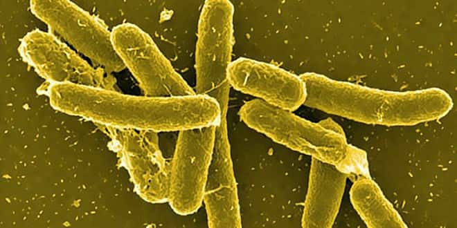
Vibrio, Aeromonas & Plesiomonas
Vibrio, Aeromonas & Plesiomonas
Similarities to Enterobacteriaceae
Gram-negative
Facultative anaerobes
Fermentative bacilli
Differences from Enterobacteriaceae
Polar flagella
Oxidase positive
Formerly classified together as Vibrionaceae
Primarily found in water sources
Cause gastrointestinal disease
Shown not closely related by molecular methods
General Characteristics of Vibrio, Aeromonas and Plesiomonas
Morphology & Physiology of Vibrio
Comma-shaped (vibrioid) bacilli
V. cholerae, V. parahaemolyticus, V. vulnificus are most significant human pathogens
Broad temperature & pH range for growth on media
18-37°C
pH 7.0 – 9.0 (useful for enrichment)
Grow on variety of simple media including:
MacConkey!s agar
TCBS (Thiosulfate Citrate Bile salts Sucrose) agar
V. cholerae grow without salt
Most other vibrios are halophilic
Morphology & Physiology of Vibrio
Vibrio spp. (Family Vibrionaceae) Associated with Human Disease
Vibrio spp. (including V. cholerae) grow in estuarine and marine environments worldwide
All Vibrio spp. can survive and replicate in contaminated waters with increased salinity and at temperatures of 10-30oC
Pathogenic Vibrio spp. appear to form symbiotic (?) associations with chitinous shellfish which serve as an important and only recently recognized reservoir
Asymptomatically infected humans also serve as an important reservoir in regions where cholera is endemic
Epidemiology of Vibrio spp.
Taxonomy of Vibrio cholerae
>200 serogroups based on somatic O-antigen
O1 and O139 serogroups are responsible for classic epidemic cholera
O1 serogroup subdivided into
Two biotypes: El Tor and classical (or cholerae)
Three serotypes: ogawa, inaba, hikojima
Some O1 strains do not produce cholera enterotoxin (atypical or nontoxigenic O1 V. cholerae)
Other strains are identical to O1 strains but do not agglutinate in O1 antiserum (non-cholera (NCV) or non-agglutinating(NAG) vibrios) (non-O1 V.cholerae)
Several phage types
Epidemiology of Vibrio cholerae
Cholera recognized for more than two millennia with sporadic disease and epidemics
Endemic in regions of Southern and Southeastern Asia; origin of pandemic cholera outbreaks
Generally in communities with poor sanitation
Seven pandemics (possible beginning of 8th) since 1817 attributable to increased world travel
Cholera spread by contaminated water and food
Human carriers and environmental reservoirs
Recent Cholera Pandemics
7th pandemic:
V. cholerae O1 biotype El Tor
Began in Asia in 1961
Spread to other continents in 1970s and 1980s
Spread to Peru in 1991 and then to most of South & Central America and to U.S. & Canada
By 1995 in the Americas, >106 cases; 104 dead
8th pandemic (??)
V. cholerae O139 Bengal is first non-O1 strain capable of causing epidemic cholera
Began in India in 1992 and spread to Asia, Europe and U.S.
Disease in humans previously infected with O1 strain, thus no cross-protective immunity
Pathogenesis of V.cholerae
Incubation period: 2-3 days
High infectious dose: >108 CFU
103 -105 CFU with achlorhydria or hypochlorhydria (lack of or reduced stomach acid)
Abrupt onset of vomiting and life-threatening watery diarrhea (15-20 liters/day)
As more fluid is lost, feces-streaked stool changes to rice-water stools:
Colorless
Odorless
No protein
Speckled with mucus
Pathogenesis of V.cholerae (cont.)
Cholera toxin leads to profuse loss of fluids and electrolytes (sodium, potassium, bicarbonate)
Hypokalemia (low levels of K in blood)
Cardiac arrhythmia and renal failure
Cholera toxin blocks uptake of sodium & chloride from lumen of small intestine
Death attributable to:
Hypovolemic shock (due to abnormally low volume of circulating fluid (plasma) in the body)
Metabolic acidosis (pH shifts toward acid side due to loss of bicarbonate buffering capacity)
Treatment & Prevention of V. cholerae
Untreated: 60% fatality
Treated: <1% fatality Rehydration & supportive therapy Oral Sodium chloride (3.5 g/L) Potassium chloride (1.5 g/L) Rice flour (30-80g/L) Trisodium citrate (2.9 g/L) Intravenous (IV) Doxycycline or tetracycline (Tet resistance may be developing) of secondary value Water purification, sanitation & sewage treatment Vaccines Virulence Factors Associated with Vibrio cholerae O1 and O139 Two Broad Classes of Bacterial Exotoxins Intracellular Targets: A-B dimeric (two domain) exotoxins: (prototype is diphtheria toxin of Corynebacterium diphtheriae): Bipartite structure: Binding domain (B) associated with absorption to target cell surface and transfer of active component (A) across cell membrane; once internalized, domain (A) enzymatically disrupts cell function Receptor-mediated endocytosis (host cell uptake and internalization of exotoxin) ADP-ribosylation of intracellular target host molecule Cellular Targets: Cytolytic exotoxins (usually degradative enzymes) or cytolysins: hemolysis, tissue necrosis, may be lethal when administered intravenously Cholera Toxin (A2-5B)(Vibrio cholerae) Chromosomally-encoded; Lysogenic phage conversion; Highly conserved genetic sequence Structurally & functionally similar to ETEC LT B-subunit binds to GM1 ganglioside receptors in small intestine Reduction of disulfide bond in A-subunit activates A1 fragment that ADP-ribosylates guanosine triphosphate (GTP)-binding protein (Gs) by transferring ADP-ribose from nicotinamide adenine dinucleotide (NAD) ADP-ribosylated GTP-binding protein activates adenyl cyclase leading to an increased cyclic AMP (cAMP) level and hypersecretion of fluids and electrolytes Mechanism of Action of Cholera Toxin 1 4 3 2 NOTE: In step #4, uptake of Na+ and Cl- from the lumen is also blocked. HCO3- = bicarbonate which provides buffering capacity. Mechanism of Action of Cholera Toxin Heparin-binding epidermal growth factor on heart & nerve surfaces Summary of Vibrio parahaemolyticus Infections Summary of Vibrio vulnificus Infections Virulence Factors Associated with Non-cholerae Vibrios (Kanagawa positive) Laboratory Identification of Vibrios Transport medium – Cary-Blair semi-solid agar Enrichment medium – alkaline peptone broth Vibrios survive and replicate at high pH Other organisms are killed or do not multiply Selective/differential culture medium – TCBS agar V. cholerae grow as yellow colonies Biochemical and serological tests Characteristics and Epidemiology of Aeromonas (Family Aeromonadaceae) Gram-negative facultatively anaerobic bacillus resembling members of the Enterobacteriaceae Motile species have single polar flagellum (nonmotile species apparently not associated with human disease) 16 phenospecies: Most significant human pathogens A. hydrophila, A. caviae, A. veronii biovar sobria Ubiquitous in fresh and brackish water Acquired by ingestion of or exposure to contaminated water or food Associated with gastrointestinal disease Chronic diarrhea in adults Self-limited acute, severe disease in children resembling shigellosis with blood and leukocytes in the stool 3% carriage rate Wound infections Opportunistic systemic disease in immunocompromised Putative virulence factors include: endotoxin; hemolysins; eneterotoxin; proteases; siderophores; adhesins Clinical Syndromes of Aeromonas Afimbriated Aeromonas hydrophila Nonadherent Afimbriated Bacterial Cells and Buccal Cells Adherent Fimbriated Bacterial Cells and Buccal Cells Fimbriated Aeromonas hydrophila Characteristics of Plesiomonas Formerly Plesiomonadaceae Closely related to Proteus & now classified as Enterobacteriaceae despite differences: Oxidase positive Multiple polar flagella (lophotrichous) Single species: Plesiomonas shigelloides Isolated from aquatic environment (fresh or estuarine) Acquired by ingestion of or exposure to contaminated water or seafood or by exposure to amphibians or reptiles Self-limited gastroenteritis: secretory, colitis or chronic forms Variety of uncommon extra-intestinal infections Epidemiological Features Aeromonas Plesiomonas Natural Habitat Source of Infection Fresh or brackish water Contaminated water or food Fresh or brackish water Contaminated water or food Clinical Features Diarrhea Vomiting Abdominal Cramps Fever Blood/WBCs in Stool Present Present Present Absent Absent Present Present Present Absent Present Pathogenesis Enterotoxin (??) Invasiveness Characteristics of Aeromonas and Plesiomonas Gastroenteritis REVIEW Vibrio spp. (Family Vibrionaceae) Associated with Human Disease REVIEW Vibrio spp. (including V. cholerae) grow in estuarine and marine environments worldwide All Vibrio spp. can survive and replicate in contaminated waters with increased salinity and at temperatures of 10-30oC Pathogenic Vibrio spp. appear to form symbiotic (?) associations with chitinous shellfish which serve as an important and only recently recognized reservoir Asymptomatically infected humans also serve as an important reservoir in regions where cholera is endemic Epidemiology of Vibrio spp. REVIEW Taxonomy of Vibrio cholerae >200 serogroups based on somatic O-antigen
O1 and O139 serogroups are responsible for classic epidemic cholera
O1 serogroup subdivided into
Two biotypes: El Tor and classical (or cholerae)
Three serotypes: ogawa, inaba, hikojima
Some O1 strains do not produce cholera enterotoxin (atypical or nontoxigenic O1 V. cholerae)
Other strains are identical to O1 strains but do not agglutinate in O1 antiserum (non-cholera (NCV) or non-agglutinating(NAG) vibrios) (non-O1 V.cholerae)
Several phage types
Epidemiology of Vibrio cholerae
Cholera recognized for more than two millennia with sporadic disease and epidemics
Endemic in regions of Southern and Southeastern Asia; origin of pandemic cholera outbreaks
Generally in communities with poor sanitation
Seven pandemics (possible beginning of 8th) since 1817 attributable to increased world travel
Cholera spread by contaminated water and food
Human carriers and environmental reservoirs
Summary of Vibrio cholerae Infections
Summary of Vibrio cholerae Infections (cont.)
Pathogenesis of V.cholerae (cont.)
Cholera toxin leads to profuse loss of fluids and electrolytes (sodium, potassium, bicarbonate)
Hypokalemia (low levels of K in blood)
Cardiac arrhythmia and renal failure
Cholera toxin blocks uptake of sodium & chloride from lumen of small intestine
Death attributable to:
Hypovolemic shock (due to abnormally low volume of circulating fluid (plasma) in the body)
Metabolic acidosis (pH shifts toward acid side due to loss of bicarbonate buffering capacity)
Virulence Factors Associated with Vibrio cholerae O1 and O139
Mechanism of Action of Cholera Toxin
Summary of Vibrio parahaemolyticus Infections
Summary of Vibrio vulnificus Infections
Virulence Factors Associated with Non-cholerae Vibrios
(Kanagawa positive)
Characteristics and Epidemiology of Aeromonas (Family Aeromonadaceae)
Gram-negative facultatively anaerobic bacillus resembling members of the Enterobacteriaceae
Motile species have single polar flagellum (nonmotile species apparently not associated with human disease)
16 phenospecies: Most significant human pathogens A. hydrophila, A. caviae, A. veronii biovar sobria
Ubiquitous in fresh and brackish water
Acquired by ingestion of or exposure to contaminated water or food
Associated with gastrointestinal disease
Chronic diarrhea in adults
Self-limited acute, severe disease in children resembling shigellosis with blood and leukocytes in the stool
3% carriage rate
Wound infections
Opportunistic systemic disease in immunocompromised
Putative virulence factors include: endotoxin; hemolysins; eneterotoxin; proteases; siderophores; adhesins
Clinical Syndromes of Aeromonas
Characteristics of Plesiomonas
Formerly Plesiomonadaceae
Closely related to Proteus & now classified as Enterobacteriaceae despite differences:
Oxidase positive
Multiple polar flagella (lophotrichous)
Single species: Plesiomonas shigelloides
Isolated from aquatic environment (fresh or estuarine)
Acquired by ingestion of or exposure to contaminated water or seafood or by exposure to amphibians or reptiles
Self-limited gastroenteritis: secretory, colitis or chronic forms
Variety of uncommon extra-intestinal infections
Characteristics of Aeromonas and Plesiomonas Gastroenteritis
…


