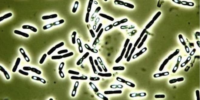
Introduction to Lab Ex. Differential Stains – Gram Staining
Introduction to Lab Ex. Differential Stains – Gram Staining
Basic classification of bacteria is based on the cell wall structure. There are 2 main groups: Gram positive and Gram negative. Gram staining is a differential staining technique that provides an easy differentiation of bacteria into one of two groups.
The staining technique, developed in the late 1700’s by Christian Gram classifies the rigid cell walled bacteria into one of two groups based on whether they are able to resist the decolorizing action of an alcoholic solution.
Those that resist decolorization by 95% ethanol are arbitrarily termed Gram positive and those that do not are Gram negative (the terms positive and negative have nothing to do with charges of the cell but based on differences in the cell wall structure of these two groups of bacteria).
The characteristic compound found in all true bacterial cell walls is peptidoglycan. The amount of PPG is among one of the differences between the GP and GN cell walls.
Gram-positive cell walls
-
Thick peptidoglycan
-
90% peptidoglycan
-
Teichoic acids
-
1 layer
-
Not many polysaccharides
-
In acid-fast cells, contains mycolic acid
Gram-negative cell
-
Thin peptidoglycan
-
5-10% peptidoglycan
-
No teichoic acids
-
3 layers
-
Outer membrane has lipids, polysaccharides
-
No acid- fast cells (mycolic acid)
…


