Enterohemorrhagic Escherichia Coli Infections
In today’s presentation we will cover information regarding Escherichia coli (E. coli) and its epidemiology. We will also talk about the history of the disease, how it is transmitted, species that it affects (including humans), and clinical and necropsy signs observed. Finally, we will address prevention and control measures for E. coli, as well as actions to take if E. coli is suspected.
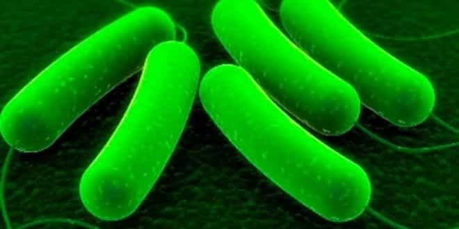
[Photo: Scanning Electron Microscopy (SEM) of Escherichia coli organism. Source: CDC Public Health Image Library]
Escherichia coli is a Gram negative rod (bacillus) in the family Enterobacteriaceae. Most E. coli are normal commensals found in the intestinal tract. Enterohemorrhagic Escherichia coli (EHEC) is a subset of pathogenic E. coli that can cause diarrhea or hemorrhagic colitis in humans. Hemorrhagic colitis occasionally progresses to hemolytic uremic syndrome (HUS), an important cause of acute renal failure in children and morbidity and mortality in adults. Pathogenic strains of this organism are distinguished from normal flora by their possession of virulence factors such as exotoxins.
[Photo: Colorized scanning electron micrograph (SEM) depicting Escherichia coli O157:H7. CDC Public Health Image Library]
The specific virulence factors can be used, together with the type of disease, to separate E. coli into pathotypes. Verocytotoxigenic (or verotoxigenic) E. coli (VTEC) produce a toxin that is lethal to cultured African green monkey kidney cells (Vero cells) but not to some other cultured cell types. There are two major families of verocytotoxins, Vt1 and Vt2. A VTEC isolate may produce one or both toxins. Because verocytotoxin is homologous to the shiga toxins of Shigella dysenteriae, VTEC are also called shiga toxin-producing E. coli (STEC). Enterohemorrhagic E. coli are VTEC that possess additional virulence factors. One key characteristic found in EHEC, but not exclusive to these organisms, is the ability to cause attaching and effacing (A/E) lesions on human intestinal epithelium. Some of the genes that are involved in producing A/E lesions can be used, together with the presence of the verocytotoxin, to help identify EHEC.
-
coli are serotyped based on the O (somatic lipopolysaccharide), H (flagellar) and K (capsular) antigens. Serotypes known to contain EHEC include E. coli O157:H7, the non-motile organism E. coli O157:H-, and members of other serogroups, particularly O26, O103, O111 and O145 but also O91, O104, O113, O117, O118, O121, O128 and others. Serotyping alone is not enough to identify an organism as an EHEC; virulence factors characteristic of these organisms must also be present. E. coli O157:H7 strains are relatively homogeneous, and nearly all of these organisms carry virulence factors associated with hemorrhagic colitis and HUS.
[Photo: Transmission electron micrograph of E. coli O157:H7 showing flagella. CDC Public Health Image Library]
-
coli O157:H7 was first described in 1982 in four patients with bloody diarrhea. The initial outbreak was associated with two outlets of the same fast-food chain, and illness was linked to undercooked hamburgers. More recently, other sources for E. coli 0157:H7 have been identified, including apple juice and cider; raw vegetables such as lettuce and spinach; raw milk; and processed foods such as salami. Over the years, E. coli O157:H7 has evolved as a major problem for physicians, public health authorities, and the food industry. Source: Centers for Disease Control and Prevention (CDC). Isolation of E. coli O157:H7 from sporadic cases of hemorrhagic colitis–United States. 1982. MMWR Morb Mortal Wkly Rep. 1997 Aug 1;46(30):700-4.
EHEC O157:H7 infections occur worldwide; infections have been reported on every continent except Antarctica. Other EHEC are probably also widely distributed. The importance of some serotypes may vary with the geographic area.
EHEC infections can occur as sporadic cases or in outbreaks. In North America, EHEC O157:H7 infections are most common from summer to autumn. Seasonality might be caused by seasonal shedding patterns in animals, or it could be due to other factors such as eating undercooked meat at summer barbecues. The incidence of EHEC in humans is difficult to determine, because cases of uncomplicated diarrhea may not be tested for these organisms. FoodNet surveillance from 1996 to 2010 showed that O157 infection caused 0.9 illnesses per 100,000; this represents a decrease compared to the period from 1996 to 1998. In clinical cases, the mortality rate varies with the syndrome. Hemorrhagic colitis alone is usually self–limiting, but death is possible. The number of cases that progress to HUS varies with the organism and the outbreak. Approximately 5-10% of patients with hemorrhagic colitis from EHEC O157:H7 usually develop HUS. Complications and fatalities are particularly common among children, the elderly, and those who are immunosuppressed or have debilitating illnesses. HUS is fatal in 3–10% of children and TTP in up to 50% of the elderly.
This image shows the relative rates of laboratory-confirmed infections with Campylobacter, STEC O157, Listeria, Salmonella, and Vibrio, compared with 1996–1998 rates, by year, according to FoodNet surveillance in the U.S. from 1996-2010. For STEC O157, a 44% decrease was observed. Compared with 2006-2008, the incidence was significantly lower for STEC O157 (29% decrease) in 2010.
Source: Centers for Disease Control and Prevention (CDC). Vital signs: incidence and trends of infection with pathogens transmitted commonly through food–foodborne diseases active surveillance network, 10 U.S. sites, 1996-2010. MMWR Morb Mortal Wkly Rep. 2011 Jun 10;60(22):749-55.
Surveys suggest that EHEC O157:H7 is widespread in cattle herds, but the prevalence in individual animals is low. Some studies have found that this organism is more common in cattle during the summer and early autumn. One study reported that the prevalence was higher when it was cooler, but more bacteria were shed in the summer. Other studies have not found seasonal patterns of shedding. Prevalence rates for EHEC O157:H7 among cattle vary from less than 1% to 36%, depending on the country, type of herd studied and other conditions. Recent studies that use sensitive methods for detection report a higher prevalence than early surveys. However, highly sensitive techniques may also overestimate prevalence, as some animals shedding the organism may not be colonized, but only transiently infected by transmission from super-shedders or the environment.
[Photo: Cattle. Source: USDA ARS]
EHEC are transmitted by the fecal–oral route. They can be spread between animals by direct contact or via water troughs, shared feed, contaminated pastures or other environmental sources. Birds and flies are potential vectors. In one experiment, EHEC O157:H7 was transmitted in aerosols when the distance between pigs was at least 10 feet. The organism was thought to have become aerosolized during high pressure washing of pens, but normal feeding and rooting behavior may have also contributed.
[Photo: Cattle at feed bunk. Source: Scott Bauer/USDA ARS]
The reservoir hosts and epidemiology may vary with the organism. Ruminants, particularly cattle and sheep, are the most important reservoir hosts for EHEC O157:H7. A small proportion of the cattle in a herd can be responsible for shedding more than 95% of the organisms. These animals, which are called super-shedders, are colonized at the terminal rectum, and can remain infected much longer than other cattle. Super-shedders might also occur among sheep. Animals that are not normal reservoir hosts for EHEC O157:H7 may serve as secondary reservoirs after contact with ruminants. Person-to-person transmission can contribute to disease spread during outbreaks; however, humans do not appear to be a maintenance host for this organism.
[Photo: Cow. Source: Larry Rana/USDA]
Foodborne outbreaks with EHEC O157:H7 are often caused by eating undercooked or unpasteurized animal products, particularly ground beef but also other meats and sausages, and unpasteurized milk and cheese. Other outbreaks have been linked to alfalfa or radish sprouts, lettuce, spinach and other contaminated vegetables, as well as unpasteurized cider. Irrigation water contaminated with feces is an important source of EHEC O157:H7 on vegetables. This organism can attach to plants, and survives well on the surface of a variety of fruits, vegetables and fresh culinary herbs. Depending on the environmental conditions, small numbers of bacteria left on washed vegetables may multiply significantly over several days. EHEC O157:H7 can be internalized in the tissues of some plants including lettuce, where it may not be susceptible to washing.
[Photos: (Top) Hamburgers. Source: USDA ARS; (Bottom) Leafy greens. Source: Centers for Disease Control and Prevention]
Multistate outbreaks of foodborne E. coli are not uncommon; for example, in recent years, the CDC has investigated food products ranging from ground beef to vegetables to prepared foods (e.g., cookie dough).
Some human cases are caused by exposure to contaminated soil or water. EHEC are usually eliminated by municipal water treatment, but these organisms may occur in private water supplies such as wells. Swimming in contaminated water, especially lakes and streams, has been associated with some infections. Soil contamination has caused outbreaks at campgrounds and other sites, often when the site had been grazed earlier by livestock.
[Photo: Small lake surrounded by farmland. Source: Neil Mitchell/geograph.org.uk (Creative Commons) via http://commons.wikimedia.org/wiki/File:Small_Lake_surrounded_by_farmland_-_geograph.org.uk_-_1752021.jpg]
The incubation period for disease caused by EHEC O157:H7 ranges from one to 16 days. Most infections become apparent after 3-4 days; however, the median incubation period was 8 days in one outbreak at an institution. Person-to-person transmission occurs by the fecal-oral route. Most people shed EHEC O157:H7 for approximately 7 to 9 days; a minority can excrete this organism for 3 weeks or longer after the onset of symptoms. In a few cases, shedding may continue for several months. Young children tend to shed the organism longer than adults. Transmission is particularly common among children still in diapers.
Most symptomatic cases begin with diarrhea. Some cases resolve without treatment in approximately a week; others progress to hemorrhagic colitis within a few days. Hemorrhagic colitis is characterized by diarrhea with profuse, visible blood, accompanied by abdominal tenderness, and in many cases, by severe abdominal cramps. Some patients have a low–grade fever; in others, fever is absent. Nausea and vomiting may be seen, and dehydration is possible. Many cases of hemorrhagic colitis are self–limiting and resolve in approximately a week. Severe colitis may result in intestinal necrosis, perforation or the development of colonic strictures.
Hemolytic uremic syndrome occurs in up to 16% of patients with hemorrhagic colitis. This syndrome is most common in children, the elderly and those who are immunocompromised. It usually develops a week after the diarrhea begins, when the patient is improving. Occasionally, children develop HUS without prodromal diarrhea. HUS is characterized by kidney failure, hemolytic anemia and thrombocytopenia. Extrarenal signs including CNS involvement with lethargy, irritability and seizures are common. In more severe cases, there may be paresis, stroke, cerebral edema or coma. Respiratory complications can include pleural effusion, fluid overload and adult respiratory distress syndrome. The form of HUS usually seen in adults, particularly the elderly, is sometimes called thrombotic thrombocytopenic purpura (TTP). In TTP, there is typically less kidney damage than in children, but neurologic signs including stroke, seizures and CNS deterioration are more common. Death occurs most often in cases with serious extrarenal disease such as severe CNS signs. Approximately 65–85% of children recover from HUS without permanent damage; however, long-term renal complications including hypertension, renal insufficiency and end-stage renal failure also occur.
Because humans do not normally carry EHEC, clinical cases can be diagnosed by finding these organisms in fecal samples. Food and environmental samples may also be tested to determine the source of the infection. There is no single technique that can be used to isolate all EHEC serotypes. Selective and differential media have been developed for EHEC O157:H7, including MacConkey agar, hemorrhagic colitis agar, and commercial chromogenic agars. Colonies suspected to be EHEC O157:H7 are confirmed to be E. coli with biochemical tests, and shown to have the O157 somatic antigen and H7 flagellar antigen with immunoassays. A variety of tests including enzyme-linked immunosorbent assays (ELISAs), agglutination, PCR, immunoblotting or Vero cell assay can be used to detect the verocytotoxin or its genes. Phage typing and pulsed field gel electrophoresis can subtype EHEC O157:H7 for epidemiology; these tests are generally done by reference laboratories. Subtyping is important in finding the source of an outbreak and tracing transmission. Serology is also valuable in humans, particularly later in the course of the disease when EHEC are difficult to find.
[Photo: MacConkey agar culture plate with Escherichia coli bacteria. Source: CDC Public Health Image Library]
Treatment of hemorrhagic colitis is supportive, and may include fluids and a bland diet. Antibiotics are controversial and are usually avoided: they do not seem to reduce symptoms, prevent complications or decrease shedding, and they may increase the risk of HUS. The use of antimotility (antidiarrheal) agents in hemorrhagic colitis also seems to increase the risk for developing HUS. Patients with complications may require intensive care including dialysis, transfusion and/or platelet infusion. Patients who develop irreversible kidney failure may need a kidney transplant.
Ruminants, especially cattle and sheep, are the major reservoirs for EHEC 0157:H7. Bison and deer can be infected. This organism can sometimes be found in other mammals including pigs, rabbits, horses, dogs, raccoons and opossums, and in birds including chickens, turkeys, geese, pigeons, gulls, rooks and various other wild birds. In some instances, it is not known whether a species normally serves as a reservoir host or if it is only a temporary carrier. For example, rabbits shedding EHEC O157:H7 have caused outbreaks in humans, but most infected rabbits have been found near farms with infected cattle. The reservoir hosts for non-O157 EHEC are poorly understood.
[Photo: (Top) Cattle. Source: Alice Welch/USDA; (Bottom) Sheep. Source: Danelle Bickett-Weddle/CFSPH]
EHEC O157:H7 has not been associated with illness in naturally infected animals. In experimentally infected calves, this serotype does not seem to cause disease in animals older than one week of age. There is one report of bloody or mucoid diarrhea, with some deaths, after experimental infection of neonatal (less than 2-day-old) calves. Another study reported illness in gnotobiotic piglets. Members of some non-O157 EHEC serogroups including O26, O111, O118 and O103 may cause diarrhea and other gastrointestinal signs in young animals. Subclinically infected animals can shed EHEC. Shedding may be transient or intermittent, and animals that have stopped excreting this organism can be recolonized. Calves are more likely to shed EHEC O157:H7 than adult cattle. Experimentally infected pigs could shed this organism for at least 2 months.
EHEC lesions in ruminants are usually characterized by inflammation of the intestinal mucosa, and are generally limited to the large intestine. In some cases, a fibrinohemorrhagic exudate is present. In rabbits experimentally infected with EHEC O153, the cecum and/or proximal colon were edematous and thickened, and the serosal surfaces had petechial or ecchymotic hemorrhages. Pale kidneys were also reported. Dogs infected with EHEC O157:H7 had no significant gross lesions. In dogs inoculated with a non-O157 EHEC strain, the primary cause of death was microvascular thrombosis leading to kidney failure and multiple organ failure. This syndrome resembled HUS. In these dogs, inflammation and edema occurred in the small and large intestines. The kidneys were pale, with a few petechiae on the serosal surface. The liver was enlarged, with inflammation and necrotic lesions.
[Photo: Hemorrhagic enteritis in canine small intestine due to E. coli O157:H7. Source: Armed Forces Institute of Pathology/CFSPH]
EHEC can be difficult to identify. They are a minor population in the fecal flora of animals. Carrier animals are usually detected by finding EHEC in fecal samples, which are either freshly voided or taken directly from the animal. Rectoanal swabs may also be used in some cases. Selective and differential media have been developed for EHEC O157:H7, including MacConkey agar, hemorrhagic colitis agar, and commercial chromogenic agars. Selective media and isolation techniques have also been developed for few non-O157 EHEC. Immunological and nucleic acid-based tests that detect O and H antigens, verocytotoxin or various genes associated with EHEC can be used for presumptive diagnosis. These rapid tests can determine whether potential pathogens are present in samples before isolation. They include dipstick and membrane technologies, agglutination tests, microplate assays, colony immunoblotting, PCR, immunofluorescence and ELISAs. Although verocytotoxin production can aid identification, VTEC are common in animals, and these organisms are not necessarily EHEC; additional virulence factors must also be identified. Verocytotoxin-negative derivatives of EHEC can occur. The results from rapid tests are confirmed by isolating the organism. Some kits validated for food and meat samples and kits for human clinical samples may lack sensitivity when testing fecal samples from animals. Although cattle can produce antibodies to O157, serology is not used routinely in animals to diagnose infections with VTEC or EHEC.
…


